Structure/Morphology
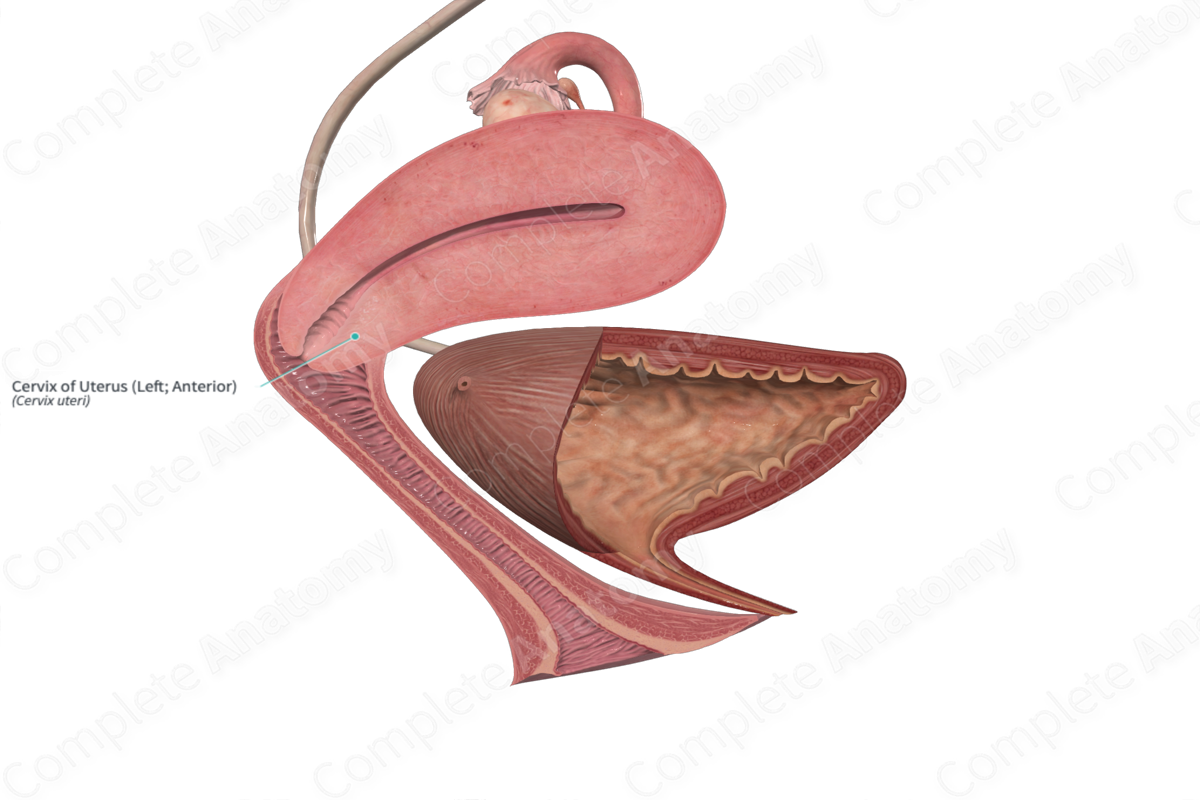
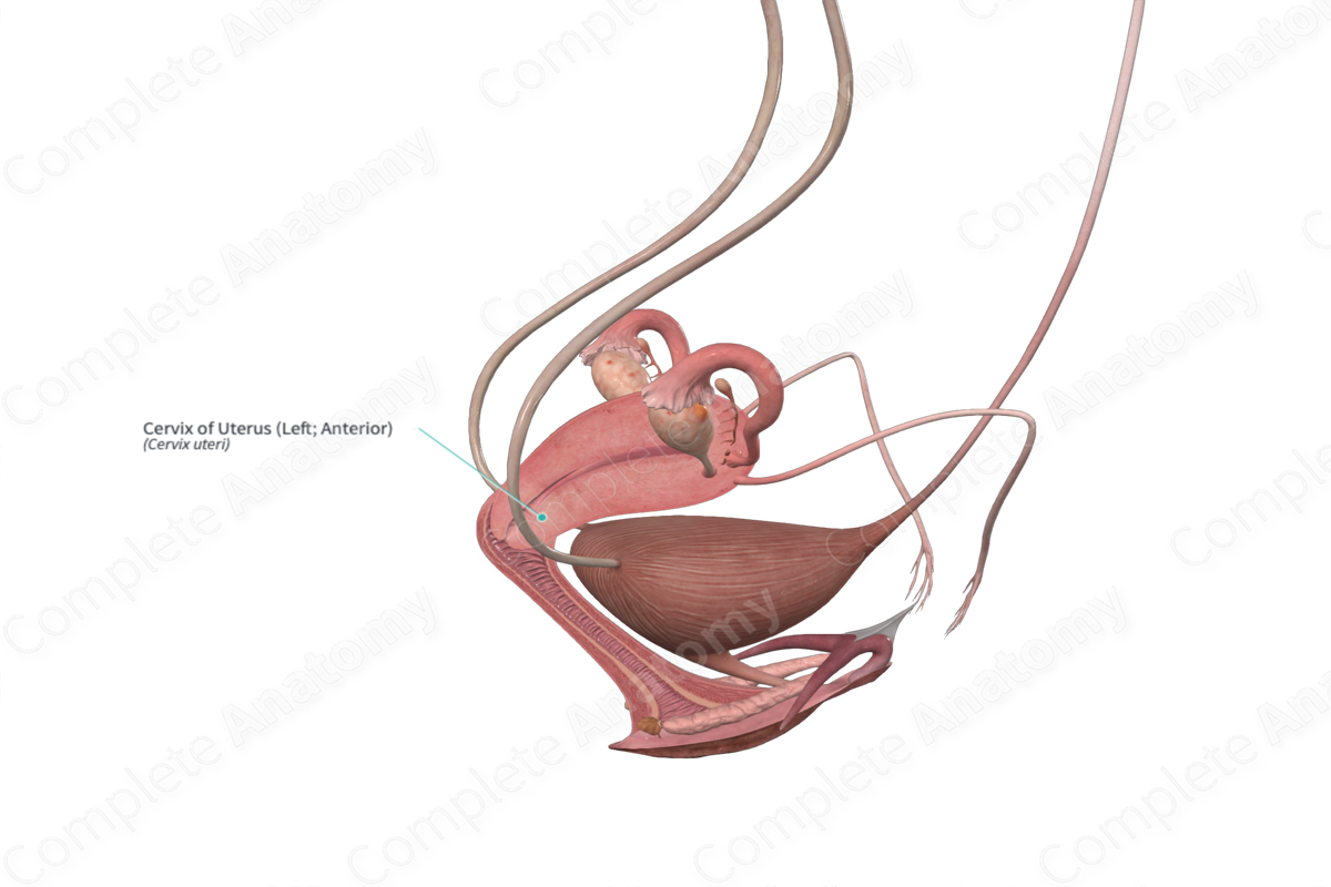
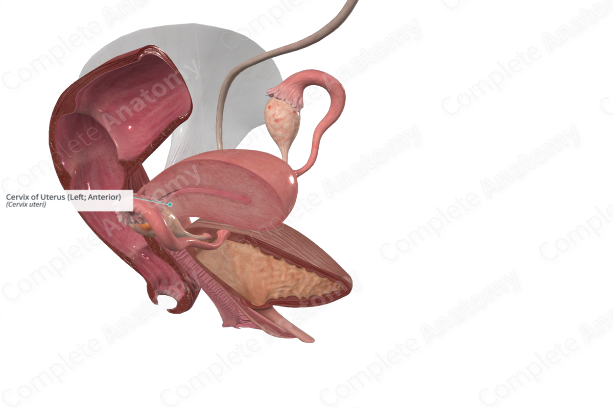
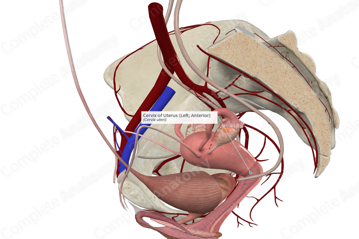
The cervix is cylindrical and approximately 2.5 cm long (Standring, 2016). The central cervical canal runs throughout the length of the cervix and connects the uterine body to the vagina. The isthmus comprises the upper one third of the cervix.
It is lined by a highly folded mucosa with columnar epithelium with tubular glands that secrete mucus. It has a fibroelastic connective tissue layer and minimum smooth muscle.
The lower portion of the cervix or ectocervix, which is at the level of the external os, is lined by a non-keratinized stratified squamous epithelium.
Key Features/Anatomical Relations
Internally, the junction between the cervix and the uterus superiorly is the internal os, while the region between the cervix and the vagina inferiorly is marked by the external os. After childbirth, the shape of the external os changes from circular to slit-like.
Both the anterior and posterior longitudinal ridges give off several folds which form a frilled edge that interdigitates to close the cervical canal.
Function
Around the time of ovulation, the cervix dilates and there is an increase in the amount of secretion, which is less viscous than previous. This facilitates sperm transportation to the site of fertilization. Additionally, the cervix dilates during parturition to facilitate the passage of the fetus through the birth canal.
List of Clinical Correlates
—Cervical cancer
—PAP smear test
—Cervical stenosis
References
Standring, S. (2016) Gray's Anatomy: The Anatomical Basis of Clinical Practice. Gray's Anatomy Series 41 edn.: Elsevier Limited.


