
Structure/Morphology
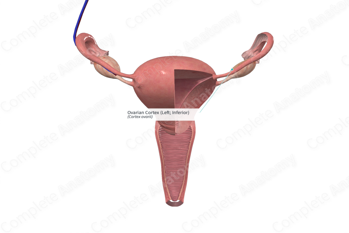
The ovarian cortex is a highly cellular stromal connective tissue, characterized by fibroblasts with collagen fibers arranged into characteristic whorls. It contains the ovarian follicles at all stages of development, both the formed and regressing corpora lutea, and the regressed corpus luteum called the corpus albicans.


Related parts of the anatomy
Key Features/Anatomical Relations
In the post-pubertal female, the cortex envelops the medulla, apart from at the hilum, and forms the bulk of the ovary. The cortex is surrounded by a thick collagenous layer called the tunica albuginea.
Function
During each menstrual cycle, up to 20 primordial follicles may develop into primary and, subsequently, secondary follicles. This growth is characterized by an increase in number and activity of the granulosa and theca cells, as well as growth of the oocyte and development of its zona pellucida.
Only one follicle becomes a tertiary (Graafian) follicle and the rest undergo atresia. Prior to ovulation, the oocyte resumes and completes meiosis I and pauses in meiosis II, where it will undergo final maturation, but only if fertilization occurs. The follicle moves to the outer region of the cortex and the stigma marks the site where the follicle bulges outwards. It’s at this point where the follicle ruptures through the tunica albuginea and, thus, where ovulation occurs.
If fertilization does not occur, the corpus luteum persists for approximately 14 days. It then undergoes luteolysis which triggers menses and the start of the next menstrual cycle. The corpus luteum continues to degenerate and after approximately two months, a whitish region of dense scar-like tissue remains in the stroma, called the corpus albicans.
If fertilization occurs, the corpus luteum persists as a hormone secreting structure throughout pregnancy and produces hormones such as progesterone and estrogen. However, the placenta takes over as the primary location for synthesis of these hormones.
List of Clinical Correlates
—Ovarian cysts
—Ovarian cancer
