Uterine Tube (Left; Anterior) Quick Facts
Location: Pelvic cavity.
Arterial Supply: Ovarian and uterine arteries.
Venous Drainage: Ovarian and uterine veins.
Innervation: Sympathetic: lumbar splanchnic nerves (T12—L2); Parasympathetic: pelvic splanchnic (S2—S4) and vagus nerves.
Lymphatic Drainage: Right, left, and intermediate lumbar lymph nodes.


Related parts of the anatomy
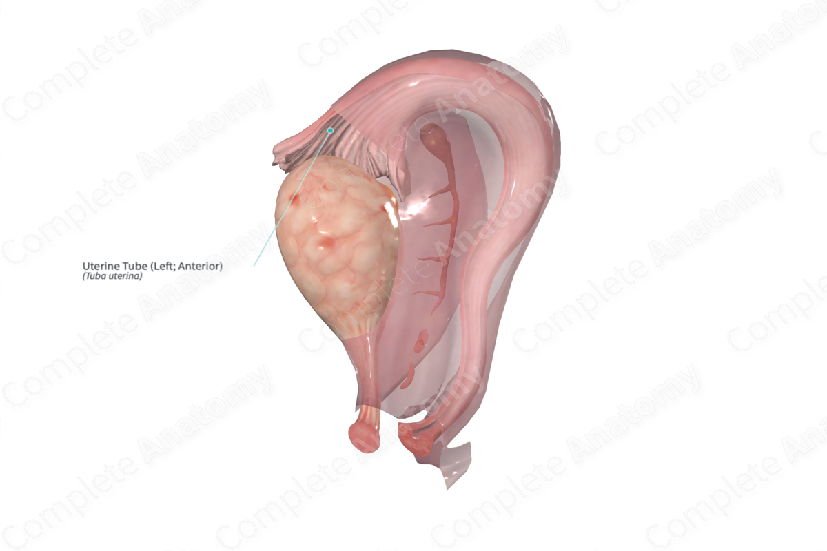
Uterine Tube (Left; Anterior) Structure/Morphology
The uterine tube is lined with an epithelial mucosa containing ciliated and secretory cells, as well as an underlying smooth muscle layer. The uterine tube can be divided into four regions, which are (from ovarian to uterine end) the infundibulum, ampulla, isthmus, and uterotubal junction.
Uterine Tube (Left; Anterior) Anatomical Relations
The uterine tube extends laterally from the superior part of the body of the uterus towards the ovary. It is enveloped within a portion of the broad ligament named the mesosalpinx.
Uterine Tube (Left; Anterior) Function
The uterine tube propels the tubal fluid and aids in the transport of the gametes and early embryo via ciliary beat and smooth muscle contraction. It contributes to the nutrition of the male and female gametes via secretion of tubal fluid. The ampulla of the uterine tube provides an appropriate environment for fertilization and is the primary location for the development and transport of the early embryo within the uterine tube.
Uterine Tube (Left; Anterior) Arterial Supply
The lateral one third is supplied by the ovarian artery and the remainder is supplied by the uterine artery.
Uterine Tube (Left; Anterior) Venous Drainage
The uterine tubes are drained via the ovarian and uterine veins. The ovarian veins drain into the inferior vena cava on the right and via the renal vein on the left.
Uterine Tube (Left; Anterior) Innervation
In order to reach the uterine tubes, nerve fibers travel through both the uterovaginal and ovarian plexuses. Sympathetic nerve supply of the uterine tubes is derived from neurons in the T10—L2 spinal segments. Parasympathetic fibers for the lateral half of the uterine tube are derived from the vagus nerve, and the medial half in supplied by the pelvic splanchnic nerves. Visceral afferent fibers travel with the sympathetic or parasympathetic fibers and enter the cord through corresponding posterior roots (Standring, 2016).
Uterine Tube (Left; Anterior) Lymphatic Drainage
Lymph from the reproductive organs such as the ovaries, uterine tube and uterus is received by the right, left, and intermediate lymph node groups (Földi et al., 2012).
Uterine Tube (Left; Anterior) List of Clinical Correlates
—Tubal ligation
—Ectopic pregnancy
Uterine Tube (Left; Anterior) References
Földi, M., Földi, E., Strößenreuther, R. and Kubik, S. (2012) Földi's Textbook of Lymphology: for Physicians and Lymphedema Therapists. Elsevier Health Sciences.
Standring, S. (2016) Gray's Anatomy: The Anatomical Basis of Clinical Practice. Gray's Anatomy Series 41 edn.: Elsevier Limited.
