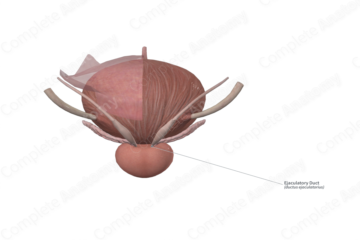
Ejaculatory Duct Quick Facts
Location: Posterior to the bladder and superior portion of the prostate gland.
Arterial Supply: Inferior vesical artery.
Venous Drainage: Prostatic and vesical venous plexuses.
Innervation: Sympathetic: Hypogastric nerve.
Lymphatic Drainage: Internal iliac lymph nodes.
Related parts of the anatomy
Ejaculatory Duct Structure/Morphology
The ejaculatory duct is a muscular tube, approximately 2 cm long, that is formed by the union of the duct of the seminal vesicle and the ampulla of the ductus deferens. Small openings of the ejaculatory duct open on both sides, or within, the prostatic utricle, to open into the prostatic urethra.
Ejaculatory Duct Anatomical Relations
The ejaculatory duct is located posterior to the bladder and superior portion of the prostate gland.
Ejaculatory Duct Function
During ejaculation, the ejaculatory duct dilates and propels sperm and seminal fluid into the prostatic urethra, and subsequently out of the body via the external urethral orifice.
Ejaculatory Duct Arterial Supply
The ejaculatory duct is supplied by the inferior vesical artery, which is a branch of the internal iliac artery.
Ejaculatory Duct Venous Drainage
The ejaculatory duct is drained by the prostatic and vesical venous plexus, which drain into the internal iliac vein.
Ejaculatory Duct Innervation
During ejaculation, the hypogastric nerve controls rapid contraction of the ejaculatory duct, by way of the sympathetic nervous system.
Ejaculatory Duct Lymphatic Drainage
The lymph of the ejaculatory duct is drained into the internal iliac lymph nodes.
Learn more about this topic from other Elsevier products
Ejaculatory Duct

The ejaculatory duct is formed by the merging of the short duct from the seminal vesicle with the vas deferens distal to the ampulla.


