Quick Facts
Location: Upper posterior abdomen.
Arterial Supply: Renal artery.
Venous Drainage: Renal vein.
Innervation: Via the renal plexus and visceral afferent fibers; Sympathetic: lowest thoracic and first lumbar splanchnic nerves, celiac and aorticorenal ganglia, aortic and celiac plexuses; Parasympathetic: posterior vagal trunk and celiac plexus.
Lymphatic Drainage: Left, right, and intermediate lumbar lymph nodes.


Related parts of the anatomy
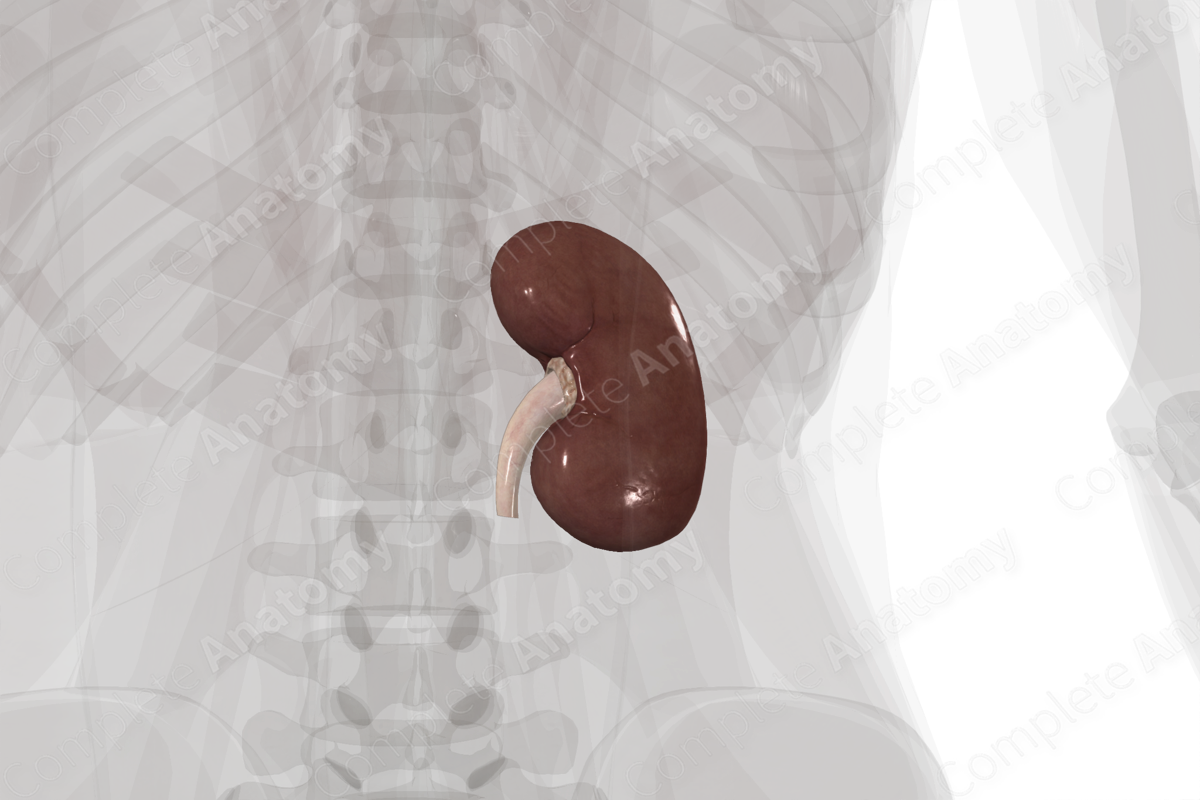
Structure/Morphology
The kidneys are bean-shaped organs with an average adult size of 11 x 6 x 3 cm (Standring, 2016). At the medial margin of the kidney, renal vessels, nerves, and a renal pelvis enter at the hilum of kidneys.
Anatomical Relations
Both the left and right kidneys sit retroperitoneal, where their posterior surfaces are embedded in fat. The left kidney is situated higher than its right counterpart. The superior pole of the left kidney sits at the level of the eleventh and twelfth ribs, while the right sits at the level of the twelfth rib (Netter, 2011). The suprarenal glands rest on the superior poles of both kidneys. The anterior surface of the right kidney is related to the right lobe of liver, descending part of duodenum, the right colic flexure, and small intestine. The anterior surface of the left kidney is related to the spleen, pancreas, stomach, left colic flexure, jejunum, and the left colic vessels.
Function
The kidneys are responsible for the excretion of liquid waste, influencing blood pressure (release of the hormone renin), red blood cell formation (release of the hormone erythropoietin), and to maintain the necessary electrolyte and water balance.
Arterial Supply
Renal arteries, originating from the abdominal aorta, enter the kidneys at the renal hilum. The arteries bifurcate into anterior and posterior branches.
Venous Drainage
The kidneys are drained by paired left and right renal veins, which drain directly into the inferior vena cava. The left renal vein is much longer than the right renal vein due to the position of the inferior vena cava on the right-hand side of the body.
Innervation
The kidneys receive visceral afferent fibers and autonomic innervation primarily through the renal plexus. Preganglionic sympathetic fibers travel primarily in the least thoracic splanchnic nerve, with variable contributions from the lesser thoracic and first lumbar splanchnic nerves. These synapse primarily on the aorticorenal ganglion but may also include the celiac and/or superior mesenteric ganglia. The parasympathetic innervation is somewhat controversial. If present parasympathetic nerves will arise from the posterior trunk of the vagus nerve, traveling through the celiac plexus (Standring, 2016).
Lymphatic Drainage
Renal lymphatic vessels remove lymph from the kidneys and send it to the right lumbar (lateral caval, precaval, and retrocaval), left lumbar (lateral aortic, preaortic and retroaortic), and intermediate lumbar lymph nodes, which return lymph to the cisterna chyli (Földi et al., 2012).
List of Clinical Correlates
—Polycystic kidney
—Horseshoe kidney
—Kidney stones
—Kidney dialysis
—End-stage renal failure
References
Földi, M., Földi, E., Strößenreuther, R. and Kubik, S. (2012) Földi's Textbook of Lymphology: for Physicians and Lymphedema Therapists. Elsevier Health Sciences.
Netter, F. H. (2011) Atlas of Human Anatomy. Netter Basic Science Series: Saunders/Elsevier.
Standring, S. (2016) Gray's Anatomy: The Anatomical Basis of Clinical Practice. Gray's Anatomy Series 41 edn.: Elsevier Limited.
Learn more about this topic from other Elsevier products
Renal, Kidney, & Urinary System Anatomy and Physiology

Review renal, kidney, and urinary system anatomy. Learn with illustrated videos and quizzes. Cover filtration, nephrons, and blood flow to prep fast.

