Structure/Morphology
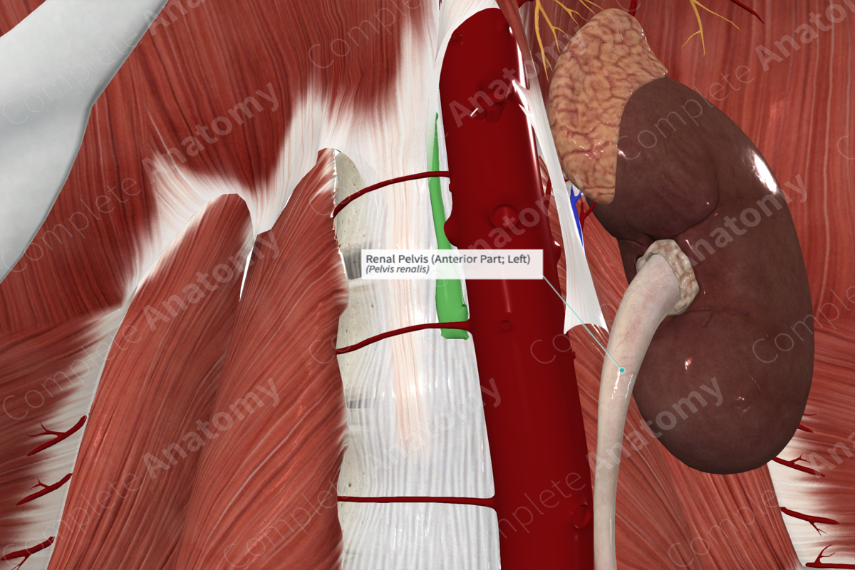
The renal pelvis is the funnel shaped, dilated proximal portion of the ureter.
Related parts of the anatomy
Key Features/Anatomical Relations
The renal pelvis is within the renal hilum, consists of a transitional epithelium (urothelium), and smooth muscle.
Function
Urine from the major calices drains into the renal pelvis before continuing distally into the ureter via peristaltic waves of smooth muscle contraction.
List of Clinical Correlates
—Kidney stones
Learn more about this topic from other Elsevier products
Renal Pelvis

Hydronephrosis is the dilation of the renal pelvis and the associated parenchymal atrophy and cystic enlargement of the kidney that results from an obstruction of urine flow.


